Drusen
| Definition | The presence of drusen is a chief characteristic of age-related macular degeneration (AMD). Unlike choroidal neovascularization (CNV) or geographic atrophy (GA) drusen can be found in early and intermediate AMD. They appear as dome-shaped structures between Bruch’s membrane and the retinal pigment epithelium (RPE) and consist mainly of lipids, but also seem to accumulate a variety of other cell components, e.g. lipofuscin or melanin. Their size is highly variable and can change over time with no clinical significance. |
|---|---|
| Clinical Symptoms | Eyes with drusen below the size of 63µm are usually not considered to suffer from AMD and the appearance is seen as a normal aging process. Drusen above 63µm have to be considered a part of early AMD. Patients with early AMD may not have any visual symptoms. Confluent large drusen in the central part of the posterior pole can reduce the visual acuity and contrast sensitivity. |
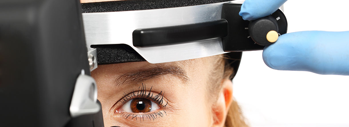
| Diagnosis and Examinations | To distinguish drusen from other retinal findings different methods of investigation are available: · Slit lamp examination · Fundus photography · Optical coherence tomography (OCT) · (Quantitative) fundus autofluorescence ((q)FAF) · Fluorescein and Indocyanine green angiography |
| Prevention and Treatment | The main and most important management for early and intermediate AMD is surveillance. Patients are advised to check regularly for metamorphopsia using the Amsler grid. General disease risk factors like smoking or obesity should be avoided. Patients should use sunglasses during sun exposure. Nutritional supplements in accordance with the AREDS2 formula is recommended. A regular clinical examination is strongly recommended to detect first signs of late AMD. |