Chorio-retinopathia centralis serosa
| Definition | Central serous chorioretinopathy (CSC) is an idiopathic disease of the retina and the choroid, which can affect one or both eyes. It can be classified in two main forms: acute and chronic. It is described as a serous detachment of the neurosensory retina and/or the retinal pigment epithelium (RPE) due to focal or diffuse leakage of fluid from the choroidal vessels. Even though the exact pathogenesis of CSC is still widely discussed and the precise pathophysiology events remain unknown, processes in stasis, ischemia or inflammation in the choroid may be responsible for the abnormal permeability and the consequent fluid accumulation. Male individuals are 6-times higher affected of CSC than female (9.9 per 100,000 to 1.7 per 100,000) and it predominantly occurs in the third and fourth decades of life. |
|---|---|
| Clinical Symptoms | The clinical symptoms of the disease can range from blurry vision combined with positive scotoma or relative scotoma (field of vision losses), metamorphopsia (blurred lines), micropsia and sometimes macropsia. Furthermore, after sunlight exposure, the recovery of the retina can be delayed. Additional the color saturation and the contrast sensitivity may be reduced. The vision acuity is normally reduced to 0.7-0.5 and can be corrected with plus lenses to 1.0. Occasional the disease appears extrafoveal and therefor asymptomatic. |
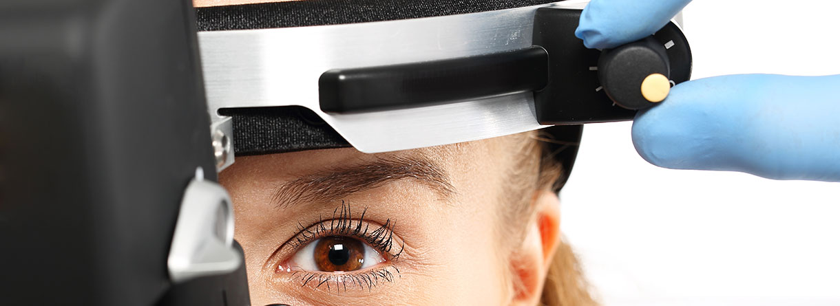
| Diagnosis and Examinations | The optical coherence tomography (OCT) and fluorescein angiography (FA) are the mainstay investigations that are helpful in confirming the clinical diagnosis, quantification of serous accumulation, documentation of pattern and site of leakage as well as in monitoring it’s recovery. Further examinations are: · Indirect ophthalmoscopy · Indocyanine green angiography (ICGA) |
| Prevention and Treatment | As the active form of CSC is a self-limited disease, with re-attachment of the neurosensory retina occurring within 3-4 months in most cases. The first-line approach consists of regular observations. There is still no consensus about the most suitable treatment and the optimal timing for interventions. Typically, treatment can be initiated if following criteria are given: persistent macular SRD for at least 4 months, history of multiple recurrences, history of CSC in the fellow eye with poor visual outcome, reduced visual acuity and a rapid recovery required. Possible treatments are exclusion of risk factors (e.g. glucocorticoid treatment and stress reduction), laser photocoagulation and verteporfin photodynamic therapy (PTD), intravitreal injections of anti-vascular endothelial growth factor (anti-VEGF) agents and medication that alter steroid hormones. |


